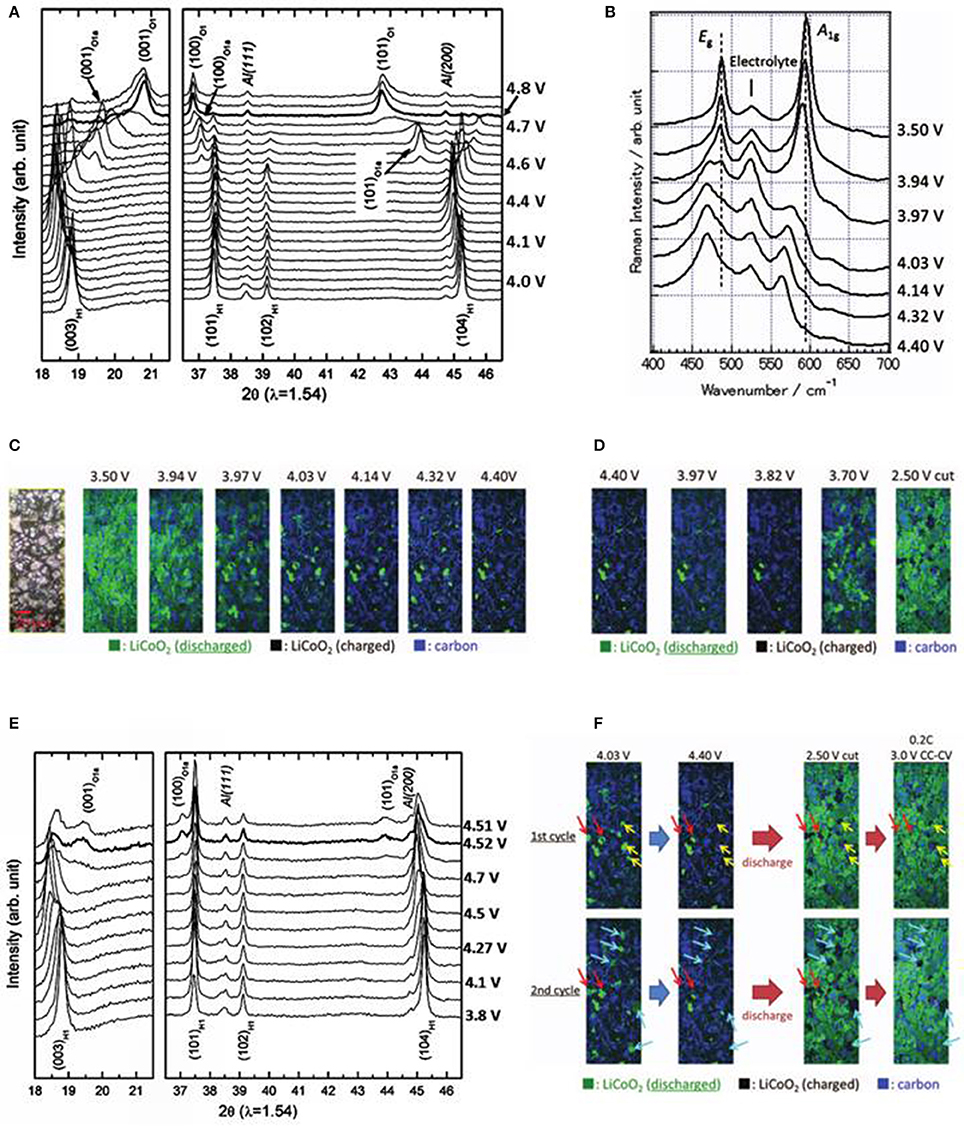
The results clearly demonstrate that in situ single-crystal X-ray diffraction is able to provide useful insights into the gradual formation of the photoproducts and the reaction processes. The work also offers a clear indication that it is possible to use the technique to study the kinetics of other photocycloaddition reactions and SCSC processes in general. An X-ray diffraction pattern is a plot of the intensity of X-rays scattered at different angles by a sample. The detector moves in a circle around the sample. The detector position is recorded as the angle 2theta (2θ) The detector records the number of X-rays observed at each angle 2θ. Energy-dispersive X-ray powder diffraction spectra have been collected for aragonite (CaC0 3) and dolomite CaMg(C0 3) 2 at high pressure and high temperature, using synchrotron radiation and a cubic multi-anvil apparatus. Unit-cell volumes were measured up to 7 GPa and 1073 K along several isothermal paths. We present in situ x-ray diffraction measurements in laser-shock compressed Fe that establish the stability of the hexagonal-close-packed (hcp) structure along the Hugoniot through shock melting, which occurs between ∼242 to ∼247 GPa.
In-situ/Operando X-ray Powder Diffraction
Overview


Real time monitoring of the formation of kinetic and thermodynamic products in a reaction is extremely important for the synthesis, isolation, and growth of new materials. Powder X-ray diffraction methods are proven to be an excellent probe for such in-situ/operando experiments . X-ray radiation of an appropriate wavelength can penetrate through the reaction vessel or device and provide diffraction information about solid-to-solid reactions, transformations, gas-to-solid interactions, liquid-to-solid crystal growth processes, and decomposition or intermediate products. Please check out some selected publications resulted by IMSERC users along with the list of other crystallographic services available in IMSERC.
User's Success Story
Applications
- Monitor reactions in real time as a function of time, temperature, pressure, and gas flow/pressure
- In-situ monitoring of crystallization processes with increasing temperature
- Operando measurements
- Accelarated aging measurements and stability tests
- Phase identification in small size samples (<1mm)
- Probe catalytic changes to substrates
If you are interested in utilizing any of these applications for your research
Selected Publications
- In-situ crystal growth using X-ray diffraction Phase Transitions in Metal–Organic Frameworks Directly Monitored through In Situ Variable Temperature Liquid-Cell Transmission Electron Microscopy and In Situ X-ray Diffraction
Lyu, Jiafei; Gong, Xinyi; Lee, Seung-Joon; Gnanasekaran, Karthikeyan; Zhang, Xuan; Wasson, Megan C.; Wang, Xingjie; Bai, Peng; Guo, Xianghai; Gianneschi, Nathan C.; Farha, Omar K. [10.1021/jacs.0c00542] - Time-Resolved in Situ Polymorphic Transformation Time-Resolved in Situ Polymorphic Transformation from One 12-Connected Zr-MOF to Another
Lee, S. J.; Mancuso, J. L.; Le, K. N.; Malliakas, C. D.; Bae, Y. S.; Hendon, C. H.; Islamoglu, T.; Farha, O. K. [10.1021/acsmaterialslett.0c00012]
Abstract
Despite extensive shock wave and static compression experiments and corresponding theoretical work, consensus on the crystal structure and the melt boundary of Fe at Earth's core conditions is lacking. We present in situ x-ray diffraction measurements in laser-shock compressed Fe that establish the stability of the hexagonal-close-packed (hcp) structure along the Hugoniot through shock melting, which occurs between ∼242 to ∼247GPa. Using previously reported hcp Fe Hugoniot temperatures, the melt temperature is estimated to be 5560(360) K at 242 GPa, consistent with several reported Fe melt curves. Extrapolation of this value suggests ∼6400K melt temperature at Earth's inner core boundary pressure.
- Received 27 July 2020
- Accepted 23 October 2020
Lg tv bluetooth setup. DOI:https://doi.org/10.1103/PhysRevLett.125.215702
© 2020 American Physical Society
